Overview
Long-term venous access is of critical importance to a wide group of patients. This is achieved by inserting tunneled lines via the internal jugular vein or the subclavian vein. This can be done either surgically or percutaneously. Combined use of ultrasonographically guided vein puncture and fluoroscopy has significantly reduced the complications related to insertion. This article gives a step-by-step guide to performing radiological insertion of a tunneled venous line via internal jugular access. In a short time, central venous access has become a very common procedure in interventional radiology. With the aid of image guidance, interventional radiologists can quickly and safely insert a variety of venous access devices in a variety of patients. Catheter malposition has been effectively eliminated and procedural-related complications such as pneumothorax have been markedly reduced.
Types of Tunneled Central Venous Catheters
There are different types of tunneled CVCs. Your doctor will talk with you about which type is best for you. A tunneled CVC may have a small cuff around the catheter. The cuff sits inside the skin tunnel. It helps to secure the catheter in place and prevent infection. The long thin hollow tubing that comes out of the skin is called a lumen. A tunneled CVC may have one lumen (single) or two or three separate lumens (double or triple lumens). A cap will cover the end of each lumen. Open-ended catheters have small clamps that close off the catheter when it is not in use. A Hickman or Apheresis catheter are types of open-ended catheters. Closed-ended catheters do not have clamps. Closed-ended catheters have a special valve at the tip of the catheter that closes it off. A Groshong is a type of closed ended catheter.
How is a Tunneled CVC Placed?
The access site is influenced by the indication and type of catheter being inserted. Nonetheless, as a general rule, the access site of choice is nearly always the right internal jugular vein. If this vein is occluded or cannot be used for some other reason, other veins in the neck may be chosen.
-
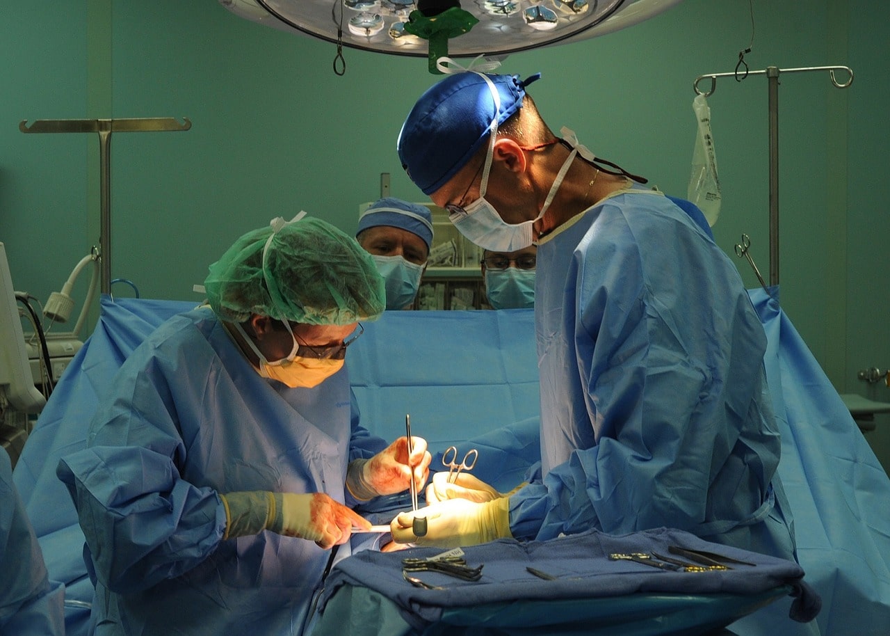
- A tunneled CVC can be put in by a doctor in the operating room or in the Radiology Department.
- An IV will be put into your arm. You will be given medicine to help you relax and you may feel drowsy.
- The skin on your neck and chest will be cleaned with a special antibacterial soap.
- A numbing medicine will be given to decrease any discomfort. This medicine will numb the skin on your chest, neck and shoulder.
- Two small incisions are made during the procedure: One incision (insertion site) will be made at the bottom of your neck near your collarbone. A second incision (exit site) will be made on your chest a few inches above your nipple. This is where the catheter comes out of your body.
- A tunnel is then made under your skin between the two incisions. The catheter will be pulled through the tunnel and then put into a large vein just above your heart. This tunneling helps to hold the catheter in place.
- The incisions will be closed and held together by stitches, special surgical glue or steri-strips (small tapes). Both incisions are covered with a small gauze dressing. The catheter line will be taped to your chest to help hold it in place.
Precision VIR is the first independent Vascular and Interventional Radiology practice in North Texas, established in 2012. We focus on minimally invasive procedures and surgeries that are the safest and most efficient for our patients.
While our website has helpful content about treatment options, we are always happy to answer any additional questions over the phone at 214-382-3200 or through an initial consultation at our Dallas clinic.